My Chart Patient Portal
 Emergency Care
 Urgent Care
 Language Services
 Medical Records
Nuclear Medicine
Nuclear medicine uses small amounts of radioactive material to diagnose and determine the severity of or treat various diseases and bodily abnormalities.
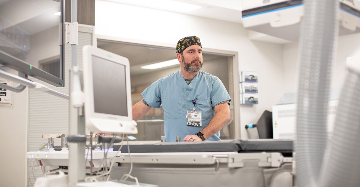
Nuclear medicine uses small amounts of radioactive material to diagnose and determine the severity of or treat various diseases and bodily abnormalities.
Nuclear medicine imaging uses small amounts of radioactive materials called radiotracers that are typically injected into the bloodstream, inhaled, or swallowed. The radiotracer travels through the area being examined. It gives off energy in the form of gamma rays which are detected by a special camera and a computer to create images of the inside of your body.
You should tell your provider if there's a possibility you are pregnant or if you are breastfeeding. Depending on the type of exam, your provider will instruct you on what you may eat or drink beforehand. You should leave jewelry at home and wear loose, comfortable clothing. You may be asked to wear a gown.
You will receive specific instructions based on the type of nuclear medicine scan you are having.
A nuclear medicine procedure uses a radioactive tracer. Most of the radioactivity passes out of your body through urine or stool. Any remaining radioactivity will disappear over time naturally.
Our staff will talk to you in detail about what you can expect before, during, and after your nuclear medicine procedure and answer any questions you may have.