My Chart Patient Portal
 Emergency Care
 Urgent Care
 Language Services
 Medical Records
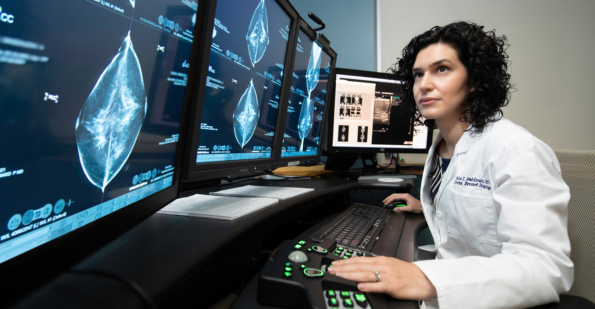
Mammography
Mammography is one of the best tools for the early detection of breast cancer. This unique x-ray can show changes in the breast up to two years before you or your doctor can feel them. Finding breast disease early greatly proves treatment options.
The American Cancer Society (ACS) recommends that women of average risk have a first, or baseline, mammogram at age 40 and annually thereafter.